
Key Takeaways
-
Educators in animal science departments and institutions across the United States face difficulty in obtaining meat carcasses for their anatomical meat science classes, impeding students' engagement in real-world immersive learning environments.
-
Distance and online learners are disadvantaged because they cannot attend hands-on laboratories to study animal carcasses and miss out on the learning that occurs from direct access to the physical meat models.
-
The University of Nebraska–Lincoln is developing an open repository of three-dimensional virtual animal carcass simulations specifically to address these access problems.
Educators in animal science departments and institutions across the United States face difficulties in obtaining meat carcasses for their anatomical meat science classes, as the carcasses are easily perishable, costly, and involve safety issues. Further, carcasses must be kept in low temperatures. As a result, carcasses are not easy to present to large groups of students because sessions must be conducted in coolers, which are typically compact, uncomfortable spaces that reach temperatures below 45º C.
Given these challenges and constraints, carcass anatomy classes are often forced to learn three-dimensional (3D) spatial relationships through 2D mediums such as textbooks and videos, ultimately impeding students' engagement in real-world immersive learning environments. In addition, even when hands-on real-world sessions are possible, distance and online learners are unable to attend and therefore fail to experience the learning that occurs in sessions with true physical meat models.
At the University of Nebraska-Lincoln, our team from the Department of Animal Science and the Nebraska Extension program set out to address these problems by developing an open repository of 3D virtual animal carcass simulations that is freely accessible to anyone. Our high-definition simulations are individually created using state-of-the art 3D scanning technologies; we then upload them to an open platform library hosted on Sketchfab, an online platform that lets people publish, share, and discover 3D and virtual reality (VR) content. Here, we describe our project and its initial results.
Creating a Simulation- and VR-Based Solution
Many universities are beginning to house multimedia equipment for faculty and staff use in spaces such as media centers and innovation labs. In this project, our team of faculty members and research assistants are using hardware available through our university's central resources service. The equipment includes a high-resolution Artec 3D scanner and a high-performance computer with a dedicated graphics card.
Scanning and Editing
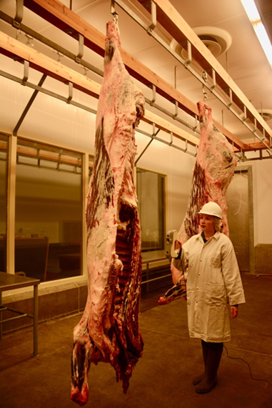
To begin this process, an operator places a portion of meat (a specimen) on a rotating table or a hook, depending on its size. The operator then scans the anatomical specimen using the Artec 3D scanner. This process captures the majority of the specimen's faces. When using a table, the Artec scanner and the accompanying Artec Studio software lets the operator capture the specimen's bottom face simply by flipping it over and resuming the scanning process. When using a hook, the user walks around the meat object while the software digitally builds a 3D model (see figure 1).
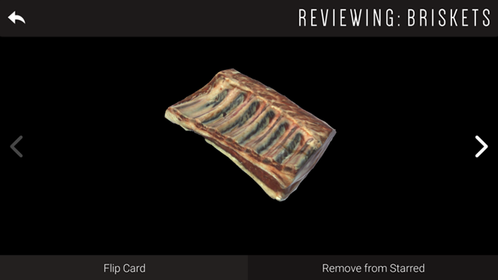
Following the basic scan procedure, Artec Studio lets us edit, texture map, and truncate any unwanted objects or (rare) errors that the scanner might capture. Having the control of post-capture processing lets us create high-resolution models that accurately represent the meat object and cut. After this post-capture processing, we upload the models to Sketchfab, where they are available for students to view using any modern Internet browser. Figure 2 shows a 3D model of a beef brisket.
Expanding Accessibility and Accuracy
As we described earlier, a major problem with current teaching methodologies is the inability to give all students a close view of dissections and anatomical breakdowns in an immersive setting. Our solution addresses this challenge.
Sketchfab integrates VR support into its platform, so students can use VR glasses to view the models more realistically. By interacting with the models using VR devices (such as Oculus, HTC Vive, or Google Cardboard) learners view a representation of a life-sized model, while also getting the full perspective and immersion of an in-person experience.
Our experiences bear this out. Through our university's 4-H extension program, we have seen how VR captures the attention of younger students and incentivizes learning in ways foreign to monolithic pedagogical techniques. 4-H youth often compete in meat-judging competitions; while recruiting young members can be difficult with traditional learning methods, the novelty and immersion that simulations offer trigger the attention of multiple age groups.1
Further, one of us (Steven Jones), often teaches introductory meat science courses over the Internet, and e-students are typically unable to visit meat lab environments. This makes it difficult for them to grasp the spatial representations of meat carcasses. Now, however, Jones' students can access and use the virtual meat models in our meat repository from their home computers. As Jones indicated, "a virtual 3D representation of meat cuts provides a dimension to carcass anatomy that has not been possible for online students in the past. This is particularly cost effective for vocational agriculture students in high school and K-12 settings."
Compared to traditional methods, which communicate information through textbooks and other (often expensive) 2D forms that are difficult to navigate, the online repository simplifies curriculum models as well. Our repository offers search and other filtration methods, giving students quick access to diverse content. For example, to access a specific anatomical object, they simply type in a specific identification number or search query to bring up the high-resolution 3D model on Sketchfab.
Clearing the Hurdles
Despite the overwhelming benefits of switching to digitizing anatomical models, some initial hurdles remain. In our case, for example, we were able to access scanning equipment and hardware through a loan from our university's central resources service. Other researchers and instructors may not have access to this equipment, which requires a hefty initial investment.
For example, the computer we used had to be equipped with a high-end graphics card and processor to ensure a fluid editing experience; the cost for this was approximately $1,500. The majority of the investment cost, however, comes from the scanner and software, which cost the university approximately $19,000.
Although upfront costs can be expensive, the long-term savings of implementing scans outweighs these costs, not least because simulations last far longer and require far less ongoing maintenance than perishable anatomical models and cadavers.
Another concern for users may be the time required to scan and catalogue the models. Fortunately, current technology is easy to use and lets you create a workflow. In a single year, our group members collaborated to easily upload more than 100 models.
Applications and Impact
Our models have begun to have an immense impact. Aside from a large audience (the models have received thousands of views), the popularity of the 3D simulations and models has inspired developers to use them to create mobile and computer applications that uniquely stimulate learners.
One example relates to 4-H competitions, in which students are tested on their knowledge of the anatomical properties of specific meat cuts. Typically, youth are unable to fully understand 3D spatial meat carcasses because they study them using 2D resources, such as posters and paper-based flashcards. To address this, our developers created a mobile application using our 3D simulations and Unity (a cross-platform game engine) that lets students study the carcasses using 3D flashcards.
Also, professors from diverse departments have expressed high levels of interest in using the 3D visualization and simulation method in other curricular forums and areas. For example, a veterinary science professor has begun the process of capturing anatomical visualizations of organs and tissues of different animals to maintain 3D structures in easy-to-manage mediums.
The technology within our workflow has high potential in other fields, especially in other anatomical forums. The goal of our educative applications is not to replace standard learning techniques and tools, but rather to create a framework for students to engage with rich media consisting of 3D models, textbooks, and images. Ideally, models such as ours will function alongside standard tools to create a synthesis of media — as well as offer many other benefits, as we now describe.
Unique Pedagogical Benefits
Our solution's benefits are clear: it increases access to and engagement with learning experiences, which in turn expands the potential for learning, both on its own and in concert with other pedagogical models.
Access and Engagement
Professors who teach in the Nebraska Extension program are asked to find methods for increasing student engagement, often through technology. The meat simulation flashcards were produced as part of this effort, with one of us (Ashu Guru) directing the team that produced this application. The application, which runs on iOS, Android, Windows, Linux, and macOS, was created for students interested in meat-judging competitions.
The expected impact of this application is immense: students unable to access laboratory resources can now learn about beef anatomy and meat cuts on their mobile devices through the 3D simulation technologies. This technology is also affordable: students who were unable to purchase expensive paper-based and textbook materials can now access the required information using an affordable mobile application (see figure 3).
Learning Modes and Models
Visualizations help make the invisible visible. Whether it is for distance students who lack access to meat carcasses or students learning the foundations of quantum models, bringing graphics to students increases their conceptual understanding.2
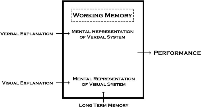
Multimodal learning spaces are key to developing long-term connections and engaging learners for longer periods of time. Integrating content from a diverse set of resources is also intrinsic to ensuring learner comprehension.3 For example, dual-coding theory4 has found that students can integrate visual and verbal information into their learning schemas more quickly than when either is presented alone (see figure 4).
Simulations enhance learning by spatially arranging components and using human sensory capacities to trigger unique perceptions of 3D configurations.5 Spatial problems, such as those in (but not limited to) the anatomical sciences, require students to construct 3D mental visualizations as a prerequisite to acting upon such information.6
As technology evolves to incorporate VR and computational reality, a new methodology of engaging readers, writers, and discussants emerges.8 In turn, as audiences and modes of expression increase, students' ability to interpret and engage with new content transforms.
In addition, this technology lets educators show physical models in class settings with numerous students, as well as offer students access to first-person models at home, building a highly functional hybrid learning space. Perhaps most importantly, virtual online simulation modeling gives educators the ability to connect to all types of students through online platforms, incorporating distance education, mobile education, and blended learning techniques.
Finally, VR gives learners the ability to control objects within their learning environments. In contrast, in traditional visualization tools, students are typically forced to navigate through what the educator wants them to see, rather than what they want to see. Having the ability to pick from an infinite number of angles to view an object gives students the choices and freedom to determine their learning pace based on their levels of cognition, spatial reasoning, recognition, and recall.
Moving Forward: Recommendations
Simulations and 3D technologies are proliferating in academia. To take full advantage of this development, we recommend that institutions take the following actions:
- Ensure that your instructional technologists and other curriculum management staff familiarize themselves with these technologies as quickly as possible.
- Train or hire staff members to specialize in handling advanced visualizations. The ease of use of modern software and hardware (such as Artec and Sketchfab) makes this an attainable goal.
- Expose students in undergraduate programs to the potential of simulation through education and workforce development.
- Give professors access to 3D scanners and other software from media centers or innovation studios so they can customize content and further enrich both their instruction and the learning that results.
Although VR has manifested in educational spaces, to date, educators themselves have had little control over its content. Enabling modular and customizable content creation lets teachers and students take advantage of visualization's potential far beyond applications in anatomical and animal sciences. Visualization technologies have proven extremely successful within educative spaces, improving both memory and learning-oriented task execution.9 For those of us interested in continuing to expand the learning horizons in higher education, mastering 3D and VR content creation opens myriad opportunities.
Notes
- Zahira Merchant, Ernest T. Goetz, Lauren Cifuentes, Wendy Keeney-Kennicutt, and Trina J. Davis, "Effectiveness of Virtual Reality-Based Instruction on Students' Learning Outcomes in K-12 and Higher Education: A Meta-Analysis," Computers & Education, Vol. 70, 2014: 29–40.
- Jong-Heon Kim, Sang-Tae Park, Heebok Lee, Keun-Cheo Yuk, and Heeman Lee, "Virtual Reality Simulations in Physics Education," Interactive Multimedia Electronic Journal of Computer-Enhanced Learning, Vol. 3, No. 2, 2001.
- Hitendra Pillay, "Cognitive Process and Strategies Employed by Children to Learn Spatial Representations," Learning and Instruction, Vol. 8, No. 1, 1998: 1-18.
- Allan Paivio and Kalman Csapo, "Picture Superiority in Free Recall: Imagery or Dual Coding?" Cognitive Psychology, Vol. 5, No. 2, 1973: 176–206.
- Wolfgang Schnotz, Emmanuel Picard, and Aemilian Hron, "How do Successful and Unsuccessful Leaners Use Texts and Graphics?" Learning and Instruction, Vol. 3, No. 3, 1993: 181–199.
- Patricia A. Carpenter and Marcel Adam Just, "Cognitive Processes in Reading," Reading Comprehension: From Theory to Practice, Lawrence Erlbaum Associates, 1986: 11–29.
- Richard E. Mayer and Valerie K. Sims, "For Whom Is a Picture Worth a Thousand Words? Extensions of a Dual-Coding Theory of Multimedia Learning," Journal of Educational Psychology, Vol. 86, No. 3, 1994: p. 389–401.
- Anne Frances Wysocki, Teaching Writing with Computers: An Introduction, 3rd ed., Houghton-Mifflin, 2002: 182–201.
- Daren T. Nicholson, Colin Chalk, W. Robert J. Funnell, and Sam J. Daniel, "Can Virtual Reality Improve Anatomy Education? A Randomised Controlled Study of a Computer-Generated Three-Dimensional Anatomical Ear Model," Medical Education, Vol. 40, No. 11, 2006: 1081–1087.
Hunter Guru is virtual reality and 3D specialist in the Department of Animal Science at the University of Nebraska–Lincoln.
Steven Jones, PhD, is a professor of muscle biology in the Department of Animal Science at the University of Nebraska–Lincoln.
Ashu Guru, PhD, is assistant professor of 4-H Youth Development at Nebraska Extension, University of Nebraska–Lincoln.
© 2017 Hunter Guru, Steven Jones, and Ashu Guru. The text of this article is licensed under Creative Commons BY-NC-ND 4.0.